
Yale scientists have successfully revived a pig brain with artificial blood
![]() 2024-08-03
2024-08-03
Recently, the cover of Nature published the latest research from Yale University School of Medicine: scientists at Yale University used an infusion solution to successfully revive the pig brain after 4 hours of death, restoring some cellular and metabolic functions, and maintaining them for at least 6 hours. It is believed that this technology will greatly benefit the development of drugs for diseases such as stroke.
The study was led by neuroscientist Nenad Sestan of the Yale School of Medicine. His team selected 32 pig brains from more than 300 pigs at a slaughter-plant in Connecticut. Using a sophisticated instrument called BrainEx, a secret formula of "artificial blood" was continuously injected into the pig brain through the artery to simulate the blood circulation in the pig brain, and the results were shocking: These pig brains can be partially "revived", some brain cells can restore basic functions, some neural activities can be reproduced, and the metabolism of the whole pig brain can become active again.

Figure 1: Sample collection site, a slaughterhouse in Connecticut
Professor Sestan said: "The original intention of this study was not to 'resurrect' the pig brain, but to try to create a more complete system to study how the brain works. Scientists used to think that if blood was cut off, the brain would die from a lack of oxygen, and unless the blood supply was quickly restored, the result was irreversible. Our assay shows that cell death is a gradual process, and some of these steps may be delayed, maintained or even reversed."
"This is a very powerful model to study the major barrier of drugs and chemicals entering the brain and penetrating the blood-brain barrier," said Yale neuroscientist Stefano Daniele, one of the lead researchers on the project. Compared with experimental animals such as mice, the pig brain is more similar to the human brain in complexity and composition, and the test results are more reliable."
It is reported that the result of this test is not a real "resurrection". The EEG measurements showed that there was no electrical activity associated with consciousness, cognition, or other higher-order brain functions throughout the trial, meaning that the pigs did not regain consciousness. In other words, the experiment only restored a certain degree of activity in the pig brain at the cellular level. 'Clinically, it's not a living brain,' says Prof Sestan. 'It's an active brain.'
The team observed that after six hours of perfusion, the pig brain was able to metabolize glucose and oxygen, a critical step in assessing cell activity. The researchers used ultrasound to look at the flow of fluid through the brain vasculature and detect hemoglobin in different areas. They added a drug to the perfusate to see how blood vessels dilated, and lipopolysaccharide, which immune cells in the brain respond to. With this, the team was able to detect the electrical signals generated by sodium and potassium ions moving in and out of hippocampal neurons to determine the cells' electrical activity.
Experts have raised concerns that the study may have ethical issues. While the team stresses that restoring consciousness is not their goal, the scientists will be monitoring the pigs' brain activity throughout the study and will act if they see any signs of a return to consciousness. Any research involving the recovery of human tissues or dead animal brains has related ethical issues and must undergo strict ethical review before proceeding.
Professor Sestan further said: "There is no EEG signal at any time in this trial. Electroencephalogram (EEG) is the recognized measure of intercellular communication related to consciousness. It is not clear whether the technique can be applied to a human brain that has not been dead for very long, because the special solution used in the trial is primarily made of biologically derived hemoglobin rather than human blood. Nevertheless, if the technology is fully developed, it will have great practical value in the future. The BrainEx system uses a cell-protective infusion that can be used to help study the structure and function of the brain in large mammals, to find out why the brain is damaged in some cases, and to test the effect of new drugs on the structure of the brain, especially for diseases like stroke, which is actually the death of part of the brain due to hypoxia."
Dennis Choi, a neuroscientist at Stony Brook University in the US, said: "This experimental model in pigs may be more suitable for simulating and developing therapies for cardiac arrest. Stroke affects only one part of the brain, whereas in cardiac arrest, hypoxia damages the whole brain, which is closer to the scenario tested."
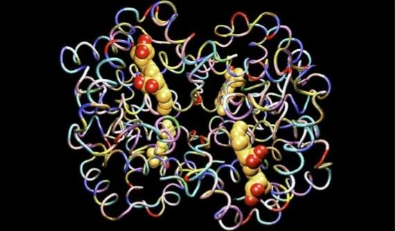
Figure 2: Oxyhemoglobin, secret formulation component of BrainEx perfusate
In the control group, the researchers used a mixture of salt and antibiotics as the perfusate. The results showed that the control group had more severe cell degeneration and apoptosis than the BrainEx group.
BrainEx perfusate is a mixture of oxygenated hemoglobin, glucose, cell death inhibitors, protein destruction inhibitors, antibiotics and neuroprotective agents. The secret ingredient is oxyhemoglobin, a substance formed by the reversibly binding of hemoglobin to oxygen molecules, which can deliver oxygen to tissues through blood flow. Its principle of action is similar to that of perfusate used in organ transplantation, which is to deliver oxygen to the whole organ tissue through oxygen carriers while regulating physiological osmotic pressure. The only difference is that the structure and physiological processes of the brain are more sophisticated than those of organs such as the liver, kidney, and heart.
This study from Yale School of Medicine also provides valuable practical experience and scientific inspiration for the application of Runfang hemoglobin oxygen supply in ex vivo organ perfusion.
-

Bios Wins the “Global Hemoglobin Oxygen Carrier Technology Innovation Leadership Award” at the Sullivan New Investment Conference 2025
On August 27, the 19th Sullivan Global Growth, Technology Innovation and Leadership Summit 2025 and the 4th New Investment Conference (hereinafter referred to as “Sullivan New Investment Conference 2025”) was grandly opened in Shangri-La Hotel, Jing ’an District, Shanghai.
2025-08-29
-

Wang Jianfeng, the Secretary of the CPC Changzhou Municipal Committee, met with Mr. Zheng Zhiheng, the Chairman of Bios Biologics
On 28 May 2025, Wang Jianfeng, Party Secretary of Changzhou City, Jiangsu Province, met with Mr. Zheng Zhiheng, Chairman of Bios Biotechnology Co., LTD., Vice Chairman and Executive Director of Chow Tai Fok Jewelry Group Co., LTD., to exchange views on further deepening cooperation.
2025-05-29
-

Yale scientists have successfully revived a pig brain with artificial blood
Recently, the cover of Nature published the latest research from Yale University School of Medicine: scientists at Yale University used an infusion solution to successfully revive the pig brain after 4 hours of death, restoring some cellular and metabolic functions, and maintaining them for at least 6 hours.
2024-08-03
-
A brief history of research and development of red blood cell substitutes
Blood has always been a very important, but very limited medical resource.
2024-08-02
